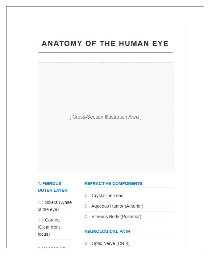
Explore the complex structure of vision with our guide on Human Eye Anatomy Layers. This resource breaks down the sclera, choroid, and retina to help students and professionals visualize ocular health. Use our Printable Chart to master identifying internal structures like the cornea, lens, and iris efficiently. Below are some ready to use templates and diagrams to assist your learning.
Image cover: Visual Guide to the Anatomical Layers of the Human Eye (Printable)
Letter Samples List
- Detailed Human Eye Anatomy Layers Printable Chart
- Labeled Human Eye Anatomy Layers Printable Chart
- Anatomical Structure Of Human Eye Layers Printable Chart
- Comprehensive Human Eye Anatomy Layers Printable Chart
- Medical Human Eye Anatomy Layers Printable Chart
- Educational Human Eye Anatomy Layers Printable Chart
- Ocular Anatomy Layers Printable Chart
- Scientific Human Eye Anatomy Layers Printable Chart
- Three Layers Of Human Eye Anatomy Printable Chart
- Internal Human Eye Anatomy Layers Printable Chart
- Biological Human Eye Layers Anatomy Printable Chart
- Detailed Anatomy Of Human Eye Layers Printable Chart
- Anatomy Of The Human Eye Layers Printable Chart
- Microscopic Human Eye Anatomy Layers Printable Chart
- Biology Study Human Eye Anatomy Layers Printable Chart
- Medical Student Human Eye Anatomy Layers Printable Chart
- Posterior Human Eye Anatomy Layers Printable Chart
- Anterior Human Eye Anatomy Layers Printable Chart
- Cross Section Human Eye Anatomy Layers Printable Chart
- Visual System Human Eye Anatomy Layers Printable Chart
Detailed Human Eye Anatomy Layers Printable Chart
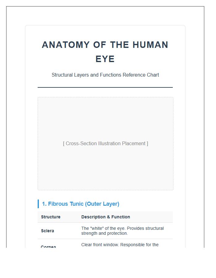
Using a detailed human eye anatomy layers printable chart is the fastest way to master complex ocular structures. This visual tool solves the problem of memorizing intricate parts like the retina and cornea. It is incredibly useful for students and medical professionals who need a clear, physical reference to simplify study sessions and improve diagnostic accuracy during patient consultations.
Labeled Human Eye Anatomy Layers Printable Chart
A Labeled Human Eye Anatomy Layers Printable Chart is an essential tool for visual learners. It simplifies complex biology by clearly identifying the retina, cornea, and iris in one view. This resource is perfect for solving study fatigue, providing a quick reference that makes mastering ocular structures both fast and effective for students or patients alike.
Anatomical Structure Of Human Eye Layers Printable Chart
Understanding vision starts with a clear visual aid. This anatomical eye chart simplifies complex layers like the retina and cornea, making it an essential tool for students or clinics. It solves the problem of visualizing internal structures, providing a quick reference to identify how light processes into sight. Download this printable resource to enhance your learning and master ocular anatomy effortlessly today.
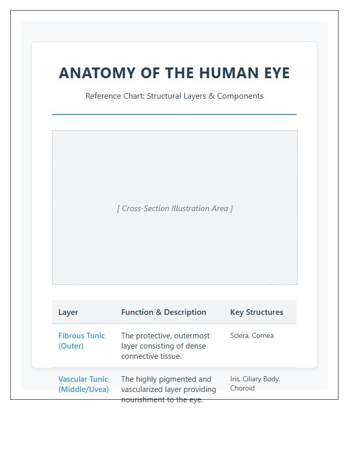
Comprehensive Human Eye Anatomy Layers Printable Chart
Understanding vision starts with a Comprehensive Human Eye Anatomy Layers Printable Chart. This visual tool is highly effective for students and professionals to master complex ocular structures. It simplifies learning by mapping each layer, solving the problem of memorizing intricate details. Having a high-quality reference makes diagnosing issues or explaining eye health much easier, ensuring you always have accurate medical information right at your fingertips.
Medical Human Eye Anatomy Layers Printable Chart
Understanding vision starts with a Medical Human Eye Anatomy Layers Printable Chart. This essential visual aid simplifies complex structures like the cornea and retina, making it perfect for students or patients. It solves the problem of memorizing intricate parts by providing a clear, high-resolution reference. Use this practical tool to enhance your learning or explain eye health clearly and effectively during consultations.
Educational Human Eye Anatomy Layers Printable Chart
An Educational Human Eye Anatomy Layers Printable Chart is an essential tool for visual learners. It simplifies complex biology by highlighting the three primary tunics and internal structures. This resource is perfect for study sessions or patient education, providing a clear, high-quality reference that solves the struggle of memorizing intricate anatomical details. Download and print it to master ocular structures with ease and precision.
Ocular Anatomy Layers Printable Chart
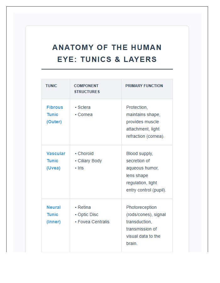
An Ocular Anatomy Layers Printable Chart is the ultimate shortcut for mastering complex eye structures. It simplifies visual learning by breaking down the fibrous, vascular, and neural tunics into clear sections. This tool is perfect for students or clinics needing a quick reference to explain conditions to patients. Having a high-quality visual makes identifying specific anatomical landmarks fast, accurate, and stress-free during study sessions.
Scientific Human Eye Anatomy Layers Printable Chart
A Scientific Human Eye Anatomy Layers Printable Chart is an essential tool for visual learners. It simplifies complex biology by clearly labeling the cornea, retina, and iris. This resource is perfect for students or clinics needing a quick reference to solve diagnostic confusion. Having a high-quality, physical diagram makes understanding vision mechanics much easier and more interactive during study sessions or patient consultations.
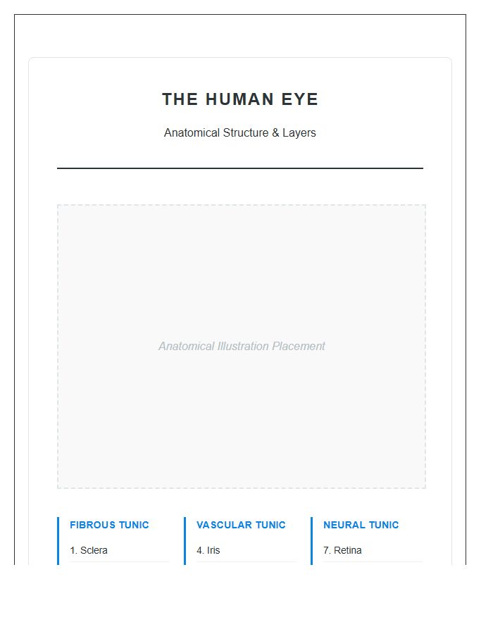
Three Layers Of Human Eye Anatomy Printable Chart
Understanding vision starts with a Three Layers Of Human Eye Anatomy Printable Chart. This visual tool simplifies complex biology, making it essential for students and health educators. It identifies the fibrous, vascular, and inner layers to help you grasp how light processes into images. Use this clear, detailed reference to solve study challenges and master ocular health concepts with total confidence and ease.
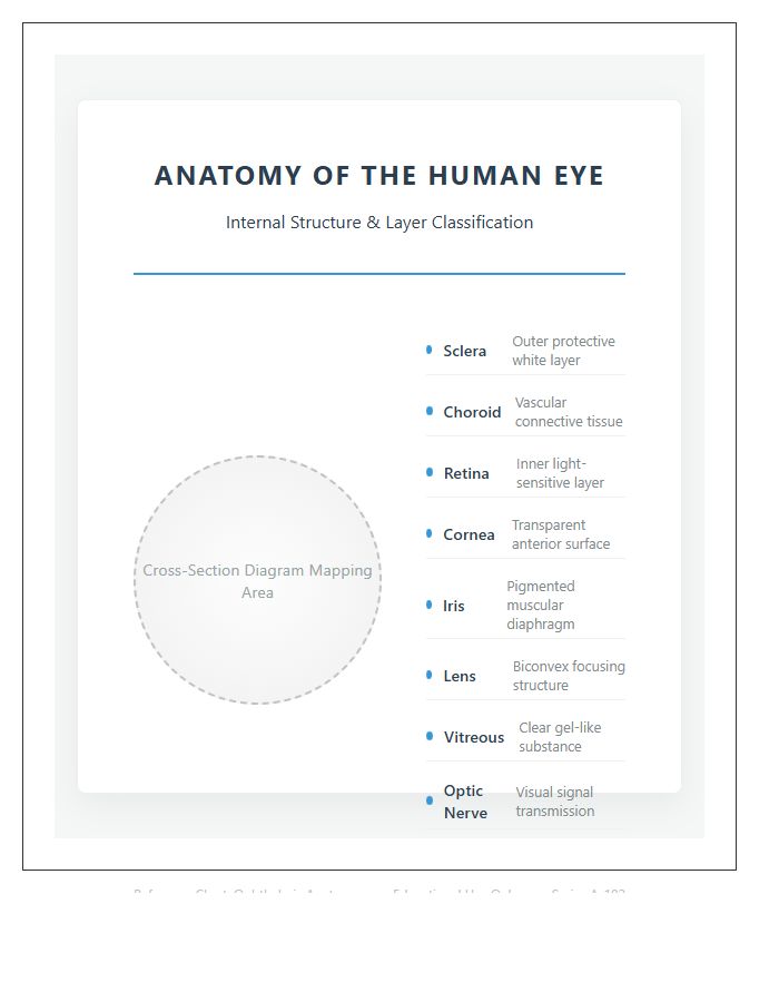
Internal Human Eye Anatomy Layers Printable Chart
An Internal Human Eye Anatomy Layers Printable Chart is an essential tool for visual learners. It simplifies complex biology by providing a clear, labeled breakdown of the ocular structure. This resource is incredibly useful for students and patients, helping them visualize how light travels through the retina and choroid. Using this chart makes understanding vision health and identifying specific anatomical parts both fast and effective.
Biological Human Eye Layers Anatomy Printable Chart
Understanding vision starts with a Biological Human Eye Layers Anatomy Printable Chart. This visual tool solves the complexity of ocular health by mapping the sclera, retina, and cornea clearly. It is incredibly useful for students and patients to grasp how light transforms into images. Having a high-quality reference ensures you master the structural details effortlessly, making it an essential resource for effective medical learning or education.
Detailed Anatomy Of Human Eye Layers Printable Chart
A detailed anatomy chart is the perfect tool for mastering complex ocular structures. This printable guide makes visualizing eye layers simple, helping students and professionals identify the cornea, retina, and iris with ease. It solves the problem of confusing diagrams by providing a clear, high-resolution reference. Use it to enhance your study sessions or explain eye health to patients effectively and quickly.
Anatomy Of The Human Eye Layers Printable Chart
Using an Anatomy Of The Human Eye Layers Printable Chart is the smartest way to simplify complex biology. This visual tool helps you master ocular structures quickly, solving the problem of confusing textbook diagrams. It is incredibly useful for students or clinics needing a clear, labeled reference to explain how vision works. Grab your high-quality chart to make learning both interactive and efficient today.
Microscopic Human Eye Anatomy Layers Printable Chart
Understanding vision starts with a Microscopic Human Eye Anatomy Layers Printable Chart. This visual tool simplifies complex structures like the retina and cornea, making it essential for students and healthcare educators. It solves the challenge of memorizing intricate ocular details by providing a clear, high-resolution reference. Whether for quick study or patient education, this chart makes mastering ocular biology both fast and remarkably easy.
Biology Study Human Eye Anatomy Layers Printable Chart
Mastering vision science is easier with a printable anatomy chart. This tool simplifies the complex three-layer structure of the human eye, helping you identify the sclera, choroid, and retina instantly. It is perfect for solving study fatigue and visualizing how light transforms into images. Using a visual guide ensures you retain key biological terms faster, making it an essential resource for any student or science enthusiast.
Medical Student Human Eye Anatomy Layers Printable Chart
Mastering vision science is easier with a Medical Student Human Eye Anatomy Layers Printable Chart. This visual tool solves the problem of memorizing complex structures by clearly labeling the sclera, choroid, and retina. It is incredibly useful for quick reviews before exams or as a handy reference during clinical rotations. Download yours today to simplify your studies and improve your diagnostic accuracy effortlessly.
Posterior Human Eye Anatomy Layers Printable Chart
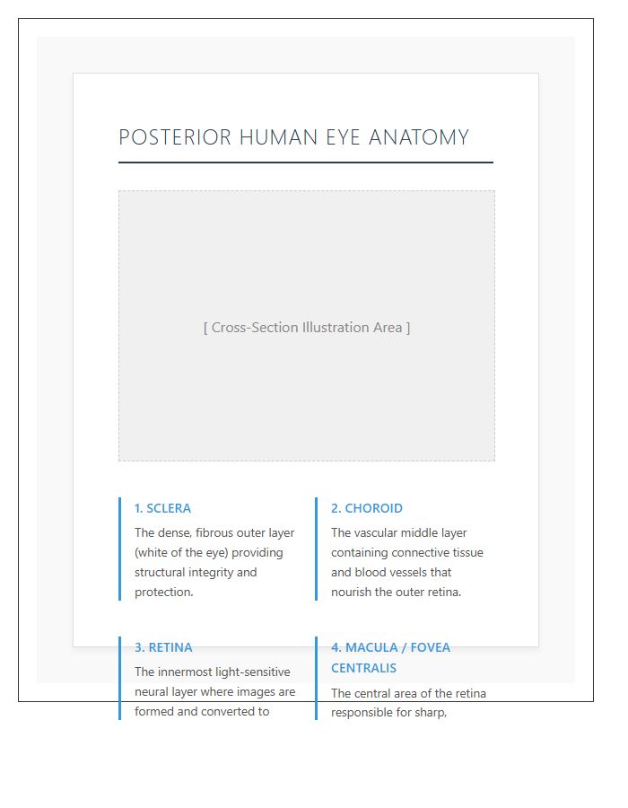
Understanding vision starts with a Posterior Human Eye Anatomy Layers Printable Chart. This essential tool simplifies complex structures like the retina and choroid for student reference. It is highly effective for visual learning, helping you master how light translates into images. Whether for clinical study or patient education, having a clear, high-quality diagram solves the problem of memorizing intricate ocular layers quickly and accurately.
Anterior Human Eye Anatomy Layers Printable Chart
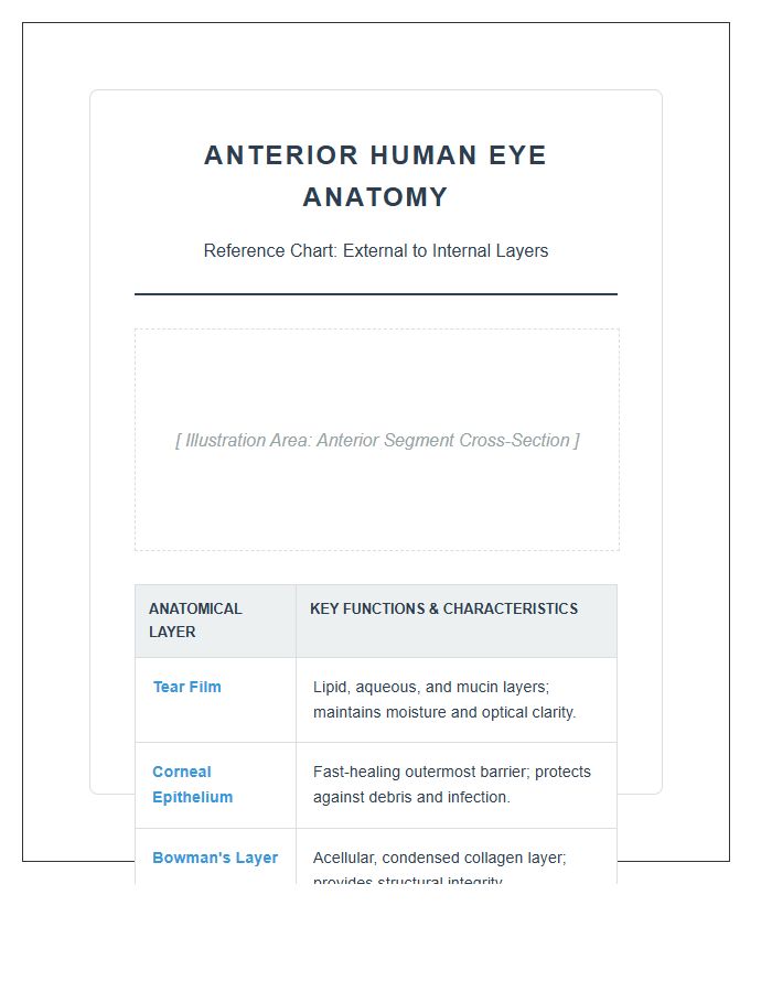
Studying vision is easier with an Anterior Human Eye Anatomy Layers Printable Chart. This visual tool solves the problem of memorizing complex structures by mapping the cornea, iris, and lens clearly. It is incredibly useful for students and educators who need a quick reference. Having a physical copy helps you master eye health concepts efficiently, making it an essential resource for medical visualization and exam preparation.
Cross Section Human Eye Anatomy Layers Printable Chart
Understanding vision starts with a Cross Section Human Eye Anatomy Chart. This printable resource simplifies complex layers like the cornea and retina, making it perfect for students or patients. It solves the problem of visualizing how light travels, providing a clear advantage for quick study sessions. Use this tool to master ocular structures and enhance your medical knowledge with ease and precision.
Visual System Human Eye Anatomy Layers Printable Chart
Understanding the human eye anatomy is essential for health education. A printable chart simplifies complex layers like the retina and cornea, making visual learning intuitive. This tool is incredibly useful for students or clinics to solve the problem of explaining vision mechanics. Having a high-quality visual guide ensures you grasp how each part functions together to create clear sight effortlessly.
What are the three primary layers of the human eye shown on a printable chart?
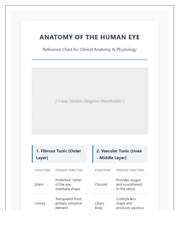
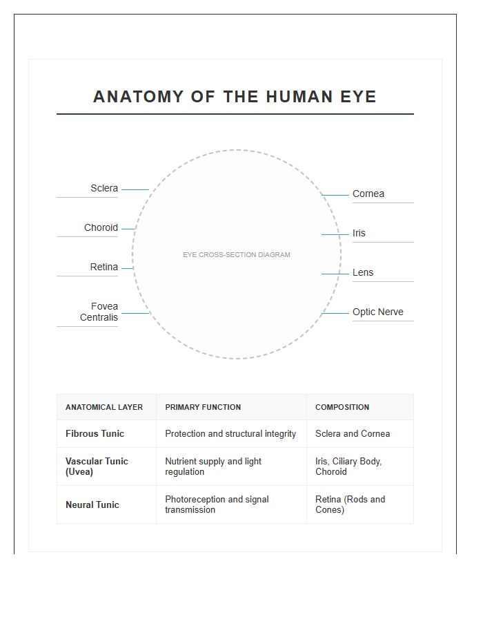
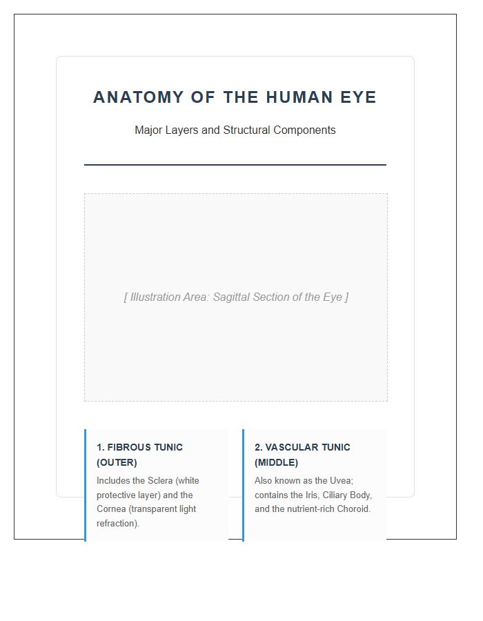
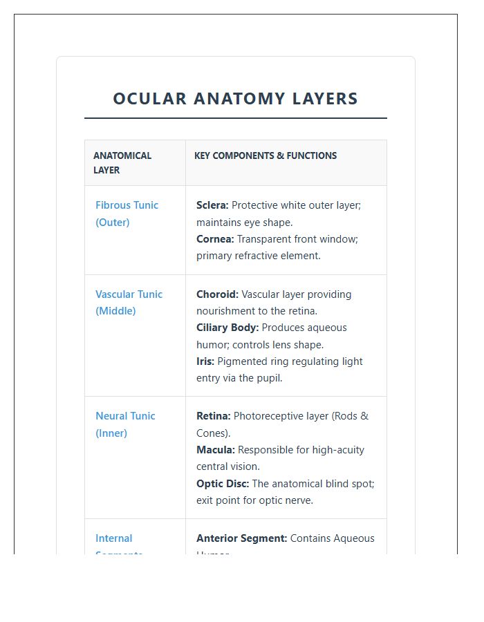
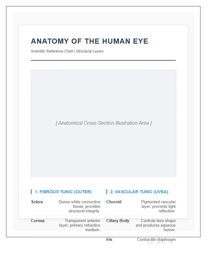
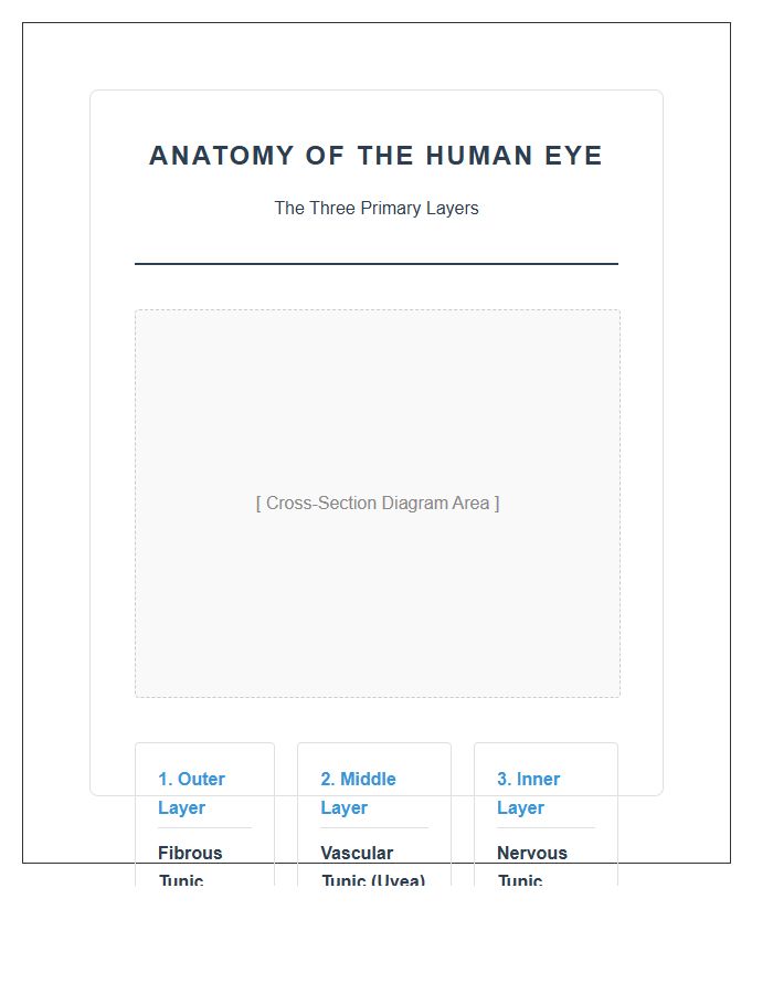
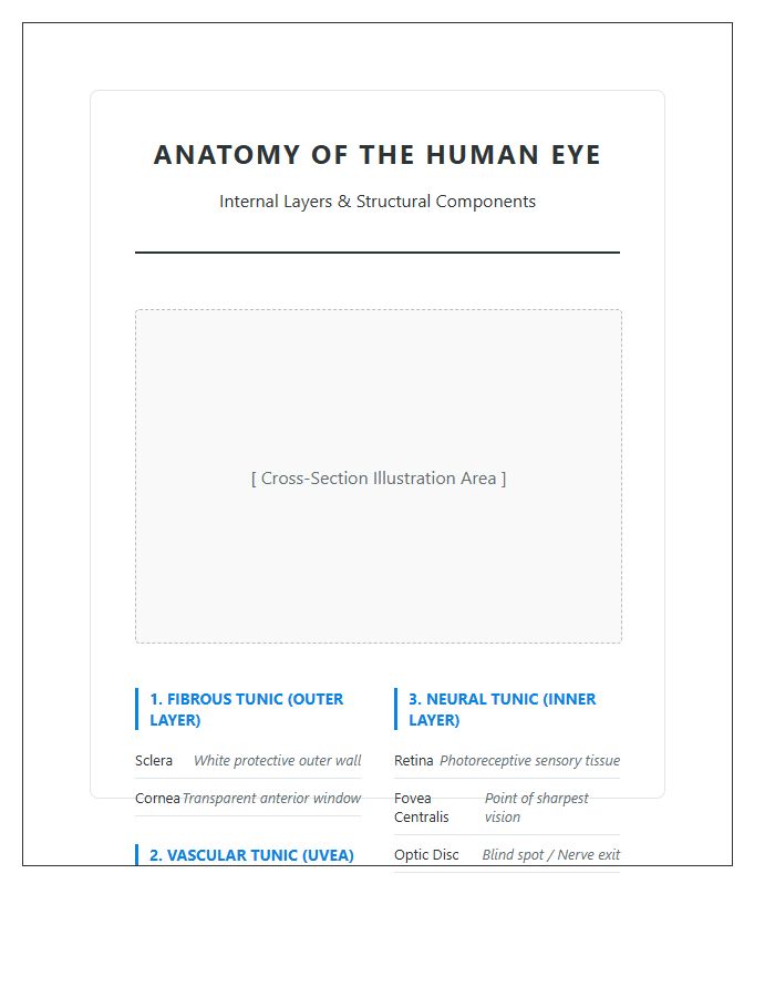
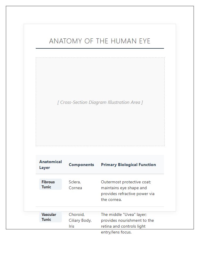
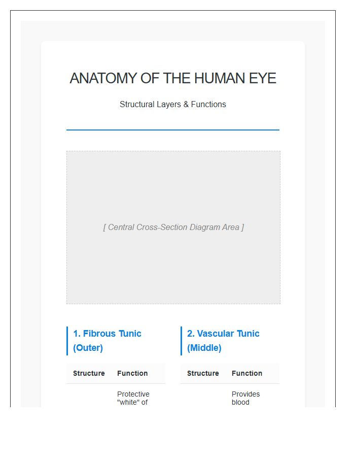
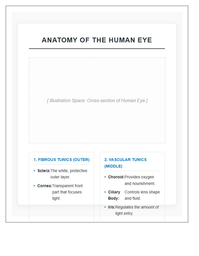
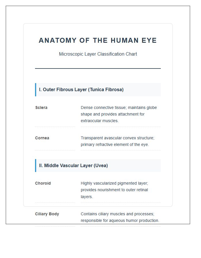
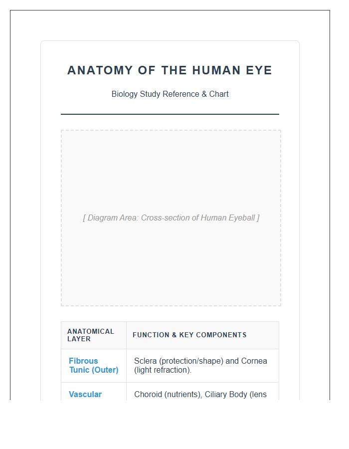
The human eye consists of three main layers: the fibrous tunic (outer layer containing the sclera and cornea), the vascular tunic or uvea (middle layer containing the iris, ciliary body, and choroid), and the neural tunic (inner layer containing the retina).
Which part of the eye anatomy is responsible for focusing light onto the retina?
Light focusing is primarily handled by the cornea and the crystalline lens. The cornea provides the majority of the eye's optical power, while the lens adjusts its shape to fine-tune focus for different distances.
Where is the retina located in a human eye anatomy diagram?
The retina is the innermost layer of the eye, lining the back of the eyeball. It contains photoreceptor cells (rods and cones) that convert light into electrical signals for the brain to interpret as images.
What is the function of the sclera in the eye's outer layer?
The sclera is the white, opaque part of the fibrous tunic that provides structural integrity, protects the inner working of the eye, and serves as an attachment point for the extrinsic muscles that move the eye.
How does a printable eye anatomy chart distinguish between the aqueous and vitreous humors?
On a printable chart, the aqueous humor is located in the anterior and posterior chambers between the cornea and lens, while the vitreous humor is the gel-like substance filling the large vitreous chamber between the lens and the retina.
Note: Sometimes the .PDF file interface can be slightly different from the image preview. Our apology for this inconvenience.

Comments