Explore the complex Large Intestine Anatomy Structure with our comprehensive guide. This anatomical overview covers the cecum, colon, rectum, and anal canal, detailing how they function in waste processing and water absorption. Perfect for students and educators, this layout clarifies segment positioning and tissue layers. Below are some ready to use printable charts to enhance your medical studies or classroom presentations.
Image cover: Visual Guide to Large Intestine Anatomy: Complete Structural Chart
Letter Samples List
- Human Large Intestine Anatomy Structure Printable Chart
- Detailed Large Intestine Anatomy Structure Printable Chart
- Large Intestine And Cecum Anatomy Printable Chart
- Large Intestine And Rectum Structure Printable Chart
- Ascending Colon Anatomy Structure Printable Chart
- Transverse Colon Anatomy Structure Printable Chart
- Descending Colon Anatomy Structure Printable Chart
- Sigmoid Colon Anatomy Structure Printable Chart
- Large Intestine Wall Layers Anatomy Printable Chart
- Large Intestine Nerve Supply Anatomy Printable Chart
- Large Intestine Blood Supply Anatomy Printable Chart
- Large Intestine Histology Structure Printable Chart
- Large Intestine Mucosa Layer Anatomy Printable Chart
- Teniae Coli Large Intestine Anatomy Printable Chart
- Haustra Of Large Intestine Anatomy Printable Chart
- Ileocecal Valve Large Intestine Anatomy Printable Chart
- Hepatic Flexure Large Intestine Anatomy Printable Chart
- Splenic Flexure Large Intestine Anatomy Printable Chart
- Large Intestine Muscularis Externa Anatomy Printable Chart
- Large Intestine Mesentery Anatomy Printable Chart
Human Large Intestine Anatomy Structure Printable Chart
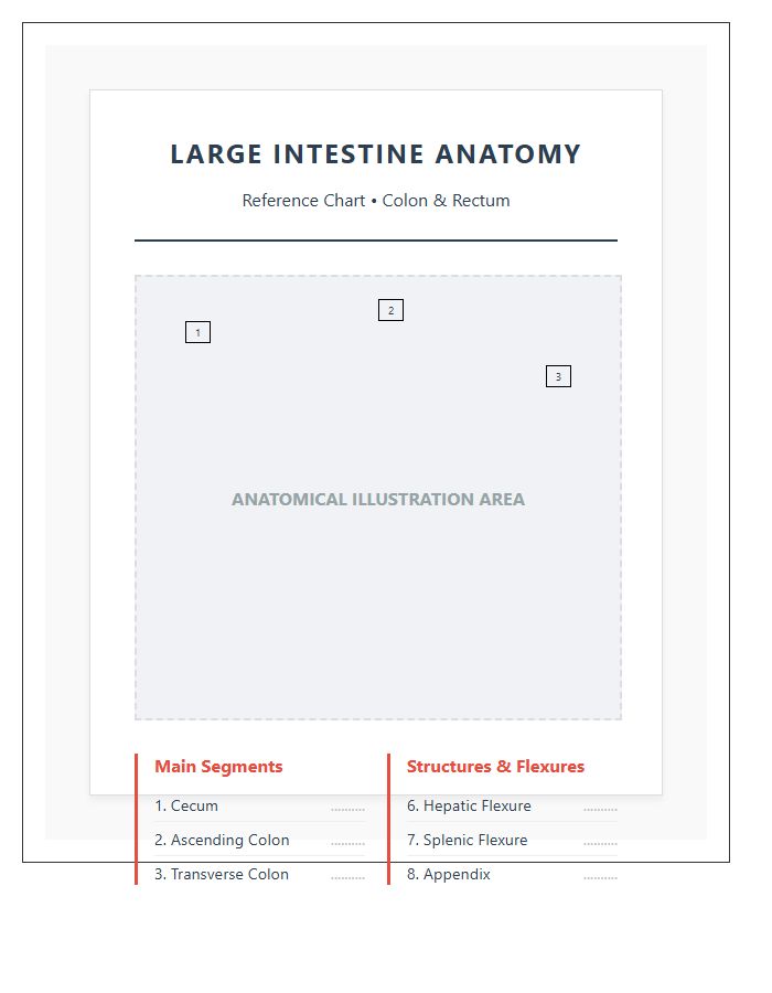
A Human Large Intestine Anatomy Structure Printable Chart is a vital tool for mastering digestive health. It simplifies complex biology, helping you visualize the colon, cecum, and rectum clearly. Whether you are a student or a patient, these diagrams solve the problem of confusing medical jargon by providing a clear, visual reference. Use it to understand nutrient absorption and improve your anatomical knowledge instantly.
Detailed Large Intestine Anatomy Structure Printable Chart
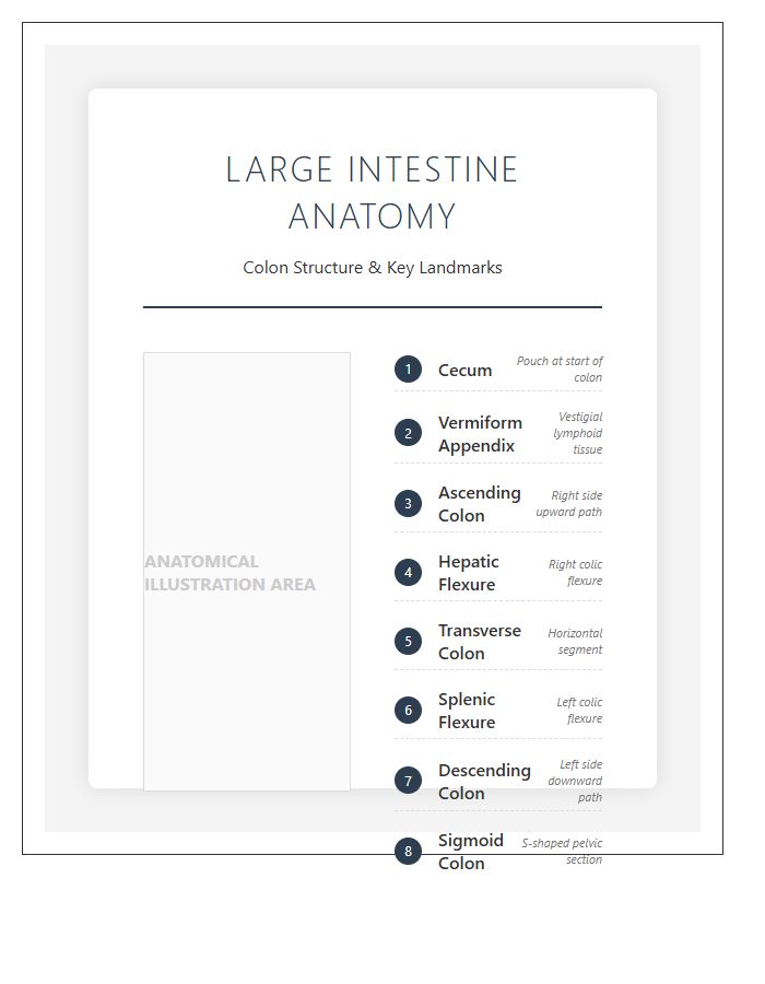
Mastering gastrointestinal health starts with clarity. This Detailed Large Intestine Anatomy Structure Printable Chart is your ultimate tool for visualizing the cecum, colon, and rectum. It solves the complexity of internal mapping, making it essential for students and clinicians. High-resolution details help you identify potential issues quickly, providing a practical advantage for both educational study and professional patient consultations.
Large Intestine And Cecum Anatomy Printable Chart
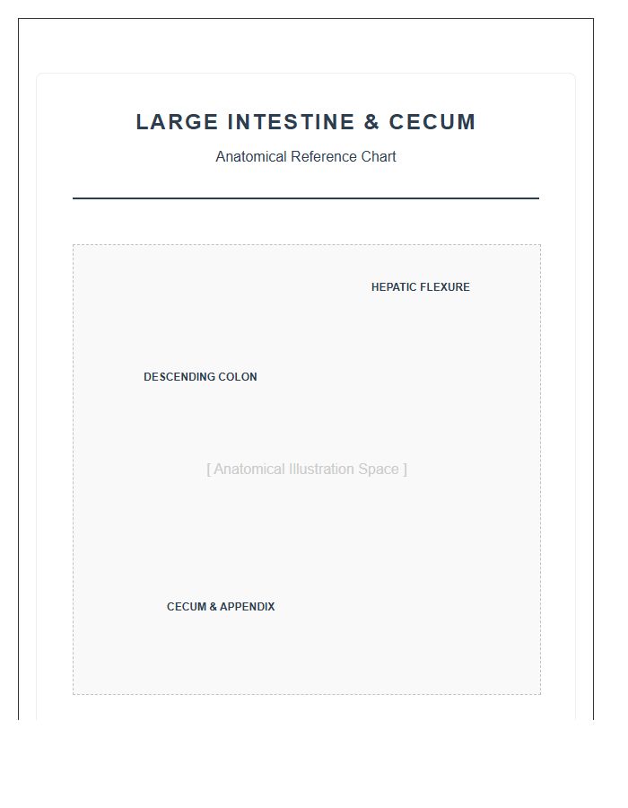
A Large Intestine And Cecum Anatomy Printable Chart is a game-changer for mastering human biology. It simplifies complex structures, making it easy to identify the ascending colon and appendix. This visual aid is perfect for students or patients needing to understand digestive health. By providing a clear, labeled overview, it solves the struggle of memorizing intestinal pathways and improves medical communication instantly.
Large Intestine And Rectum Structure Printable Chart
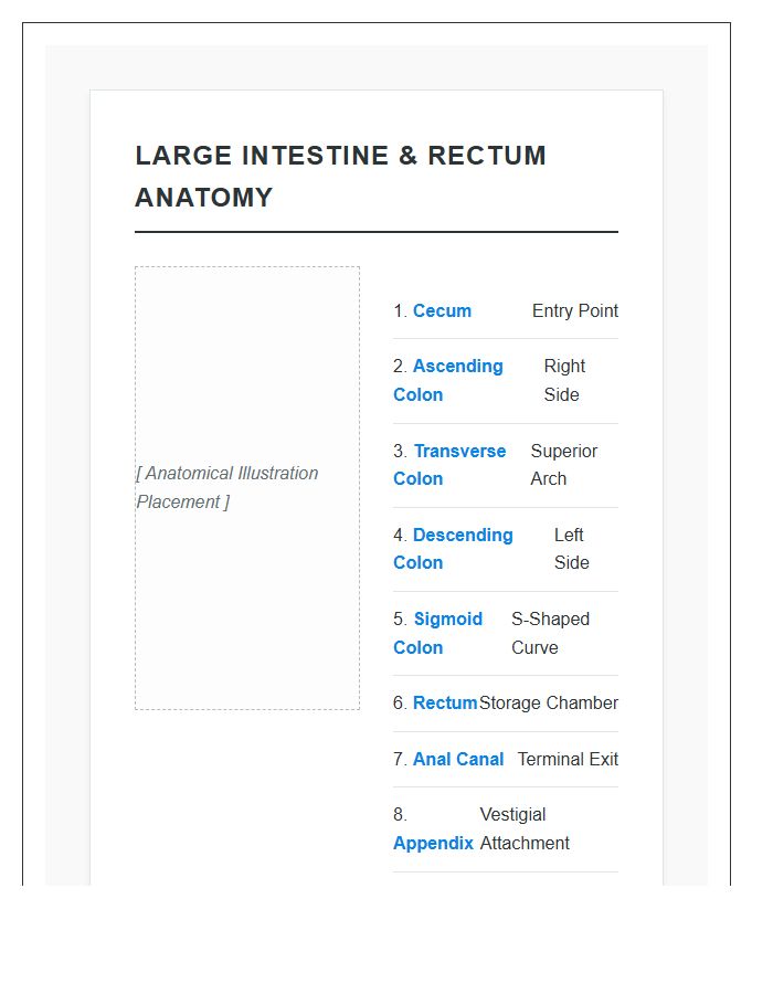
Understanding your body is easier with a Large Intestine And Rectum Structure Printable Chart. This visual tool solves the problem of complex medical jargon by providing a clear, anatomical guide to digestive health. It is incredibly useful for students or patients needing to visualize organ functions. Having this reference helps you track wellness and communicate better with doctors during check-ups.
Ascending Colon Anatomy Structure Printable Chart
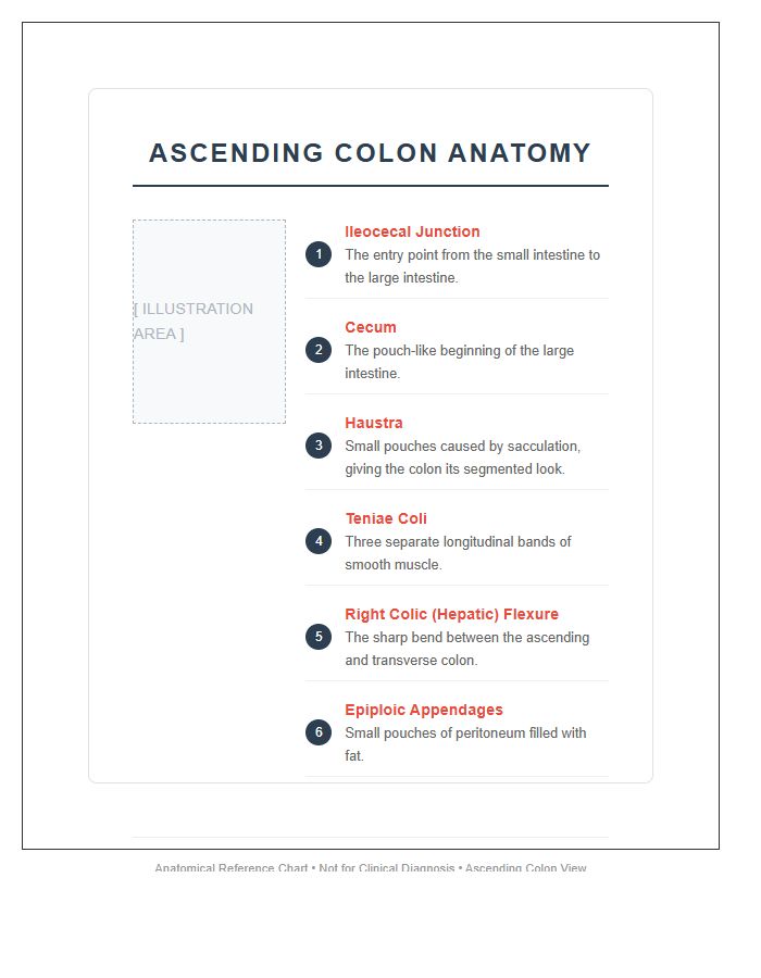
Mastering gastrointestinal health starts with clarity. An Ascending Colon Anatomy Structure Printable Chart is the perfect tool for visualizing how waste moves upward from the cecum. It simplifies complex medical terminology, making it incredibly useful for patient education or student study sessions. Having this detailed reference helps you solve anatomical confusion and quickly identify key structures in the digestive system with total confidence.
Transverse Colon Anatomy Structure Printable Chart
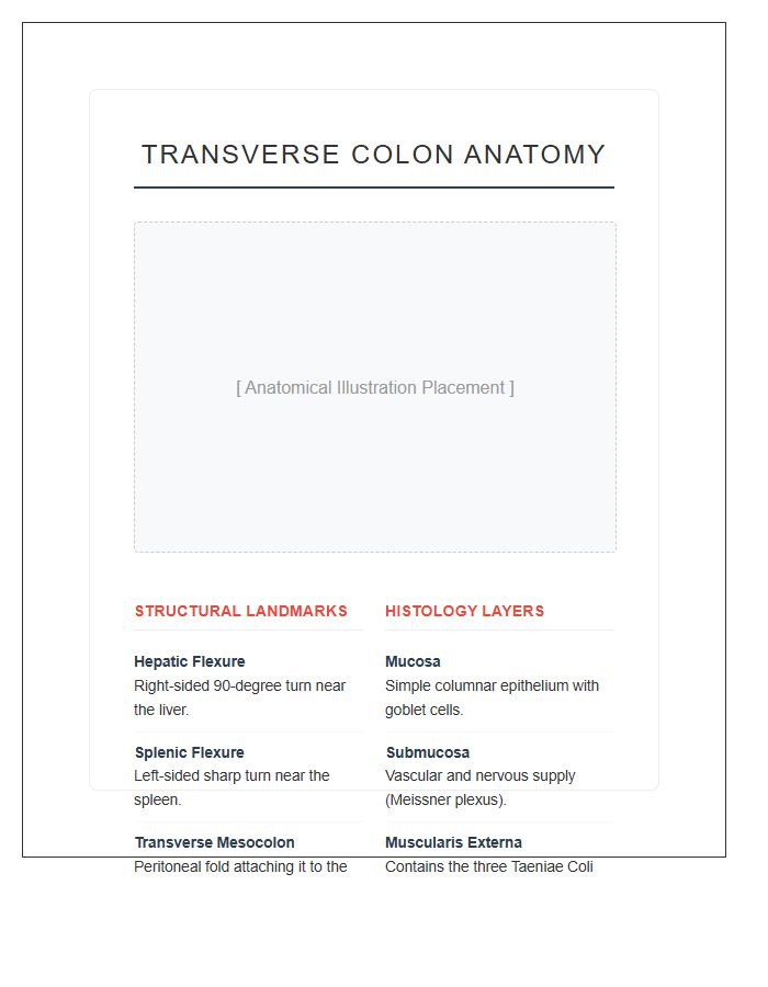
A Transverse Colon Anatomy Structure Printable Chart is a vital tool for visualizing how the large intestine crosses the abdomen. It helps students and patients understand the digestive pathway clearly. Using this chart simplifies learning complex internal organs, making it an effective resource for solving anatomical confusion. It provides a quick, portable reference that ensures you grasp the gut's layout with ease and total accuracy.
Descending Colon Anatomy Structure Printable Chart
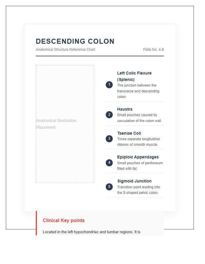
Understanding the descending colon is essential for tracking digestive health. A printable chart provides a clear visual aid for identifying its role in waste storage and water absorption. Having this anatomy structure handy is incredibly useful for patient education or studying, helping you solve complex medical concepts with simple imagery. It is a vital tool for mastering intestinal anatomy quickly and effectively.
Sigmoid Colon Anatomy Structure Printable Chart
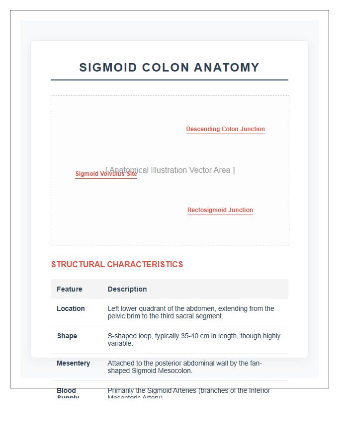
A Sigmoid Colon Anatomy Chart is an essential tool for visualizing the S-shaped curve of the lower intestine. It is incredibly useful for identifying potential issues like diverticulitis or polyps during consultations. Having a high-quality printable version solves the problem of explaining complex structures, making it easier to educate patients clearly and effectively. It is a must-have resource for both medical students and healthcare professionals.
Large Intestine Wall Layers Anatomy Printable Chart
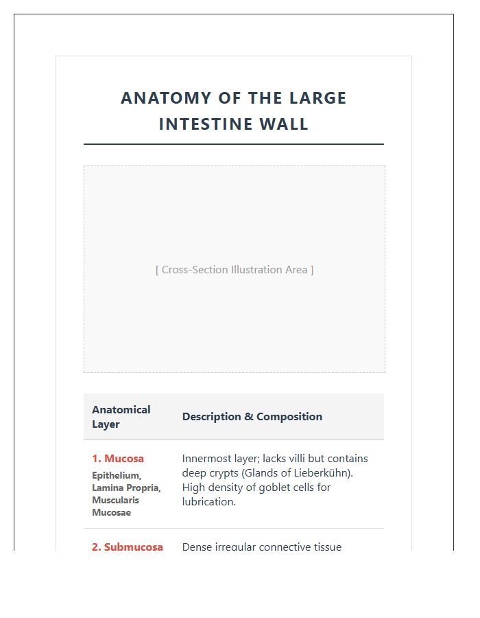
Visualizing the Large Intestine Wall Layers helps you master complex gastrointestinal anatomy effortlessly. This printable chart simplifies studying the mucosa, submucosa, and muscularis, making it an essential tool for medical students or clinicians. By clearly mapping each structural layer, you solve the problem of memorizing intricate details, providing a clear visual advantage for exams or patient education. It is practical, detailed, and highly effective.
Large Intestine Nerve Supply Anatomy Printable Chart
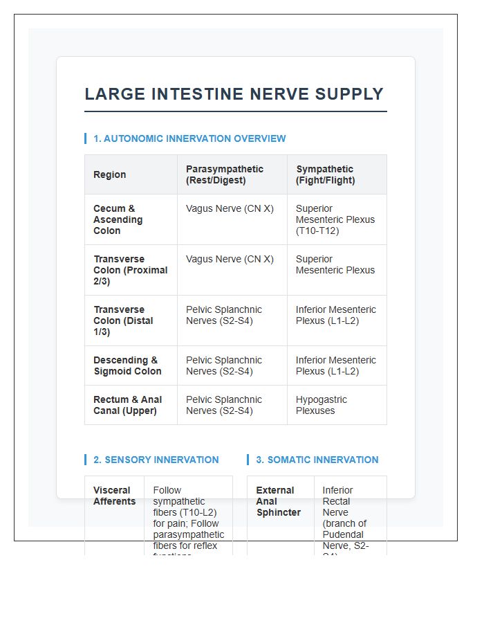
Visualizing the enteric nervous system is essential for mastering clinical assessments. Our Large Intestine Nerve Supply Anatomy Printable Chart simplifies complex sympathetic and parasympathetic pathways into clear, color-coded diagrams. This tool solves the challenge of memorizing innervation zones, helping you quickly identify the root causes of digestive dysfunction. It is a perfect reference for students and professionals seeking a practical edge in gastrointestinal studies.
Large Intestine Blood Supply Anatomy Printable Chart
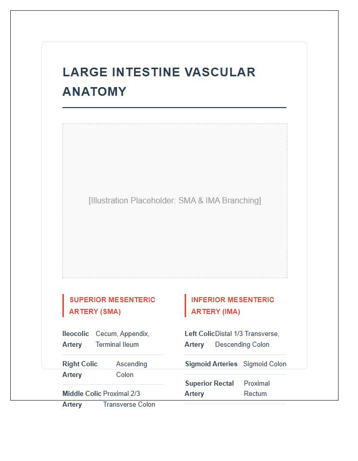
Mastering the Large Intestine Blood Supply is essential for surgical precision and clinical exams. This printable chart offers a visual advantage by mapping the mesenteric arteries and critical watersheds. It solves the complexity of vascular patterns, providing a clear reference for identifying supply zones quickly. It is an indispensable tool for anyone needing to memorize anatomical pathways for better patient outcomes or academic success.
Large Intestine Histology Structure Printable Chart
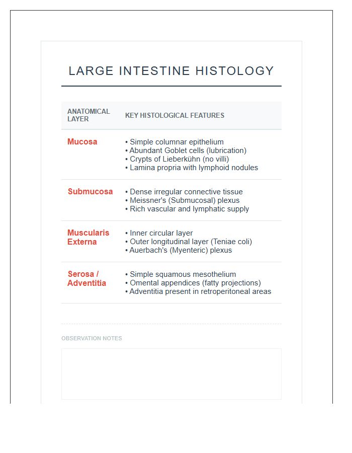
Understanding the gut's anatomy is easier with a Large Intestine Histology Structure Printable Chart. This visual tool simplifies complex layers like the mucosa and muscularis, making it essential for students mastering digestive health. It solves the struggle of memorizing cell types by providing a clear, high-resolution reference. Perfect for quick study sessions, it ensures you grasp vital structural details quickly and effectively.
Large Intestine Mucosa Layer Anatomy Printable Chart
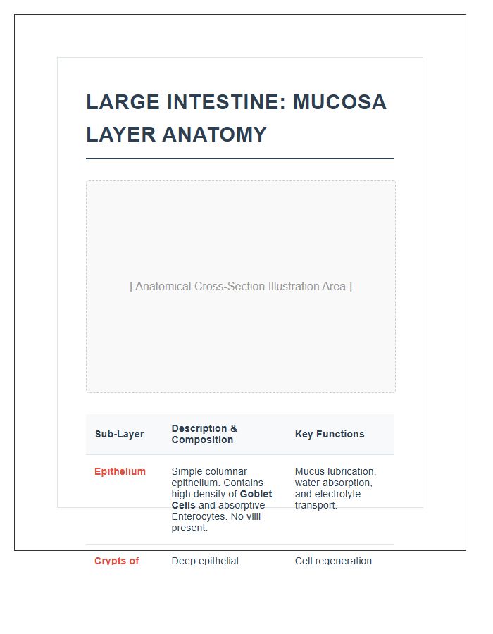
Understanding your gut health starts with clear visuals. This Large Intestine Mucosa Layer Anatomy Printable Chart is a game-changer for students and patients alike. It simplifies complex layers, helping you visualize how tissues protect your body. By using this detailed guide, you can easily identify structures and solve the mystery of digestive health, making it an essential tool for quick, effective learning and reference.
Teniae Coli Large Intestine Anatomy Printable Chart
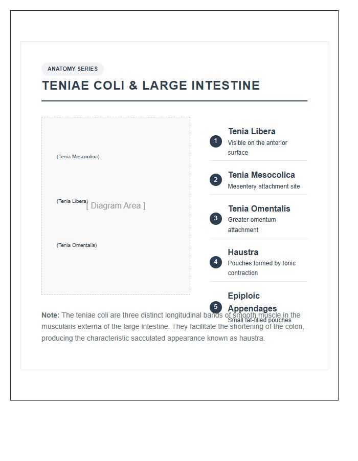
Understanding digestive health is easier with a Teniae Coli Large Intestine Anatomy Printable Chart. This visual tool helps you identify the three longitudinal muscle bands responsible for creating haustra. It is perfect for medical students or patients needing a clear explanation of bowel function. Having this high-resolution reference solves the complexity of internal structures, making study sessions or clinical consultations much more effective and professional.
Haustra Of Large Intestine Anatomy Printable Chart
Understanding the haustra of the large intestine is essential for mastering digestive anatomy. Our printable chart helps you visualize these small pouches, making it easier to identify how they facilitate segmented contractions and water absorption. It is a perfect tool for solving complex study sessions by providing a clear, labeled reference that simplifies your learning process and improves memory retention during exams.
Ileocecal Valve Large Intestine Anatomy Printable Chart
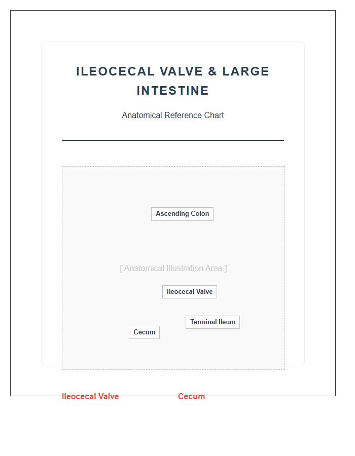
Understanding the ileocecal valve is vital for digestive health. This anatomical gateway prevents waste backflow from the large intestine to the small intestine. Our printable chart simplifies complex anatomy, helping you visualize valve function to solve issues like SIBO or bloating. It is a practical tool for patients and students to master gut health through clear, accurate visual education.
Hepatic Flexure Large Intestine Anatomy Printable Chart
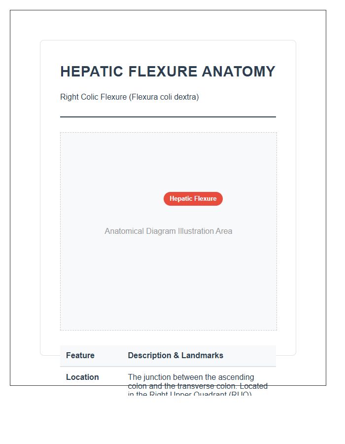
Understanding your digestive system is easier with a Hepatic Flexure Large Intestine Anatomy Printable Chart. This visual tool solves the mystery of where your ascending colon meets the transverse colon near the liver. It is a vital resource for students and patients to track digestive health, visualize potential discomfort zones, and master abdominal anatomy through clear, high-quality illustrations you can reference anytime.
Splenic Flexure Large Intestine Anatomy Printable Chart
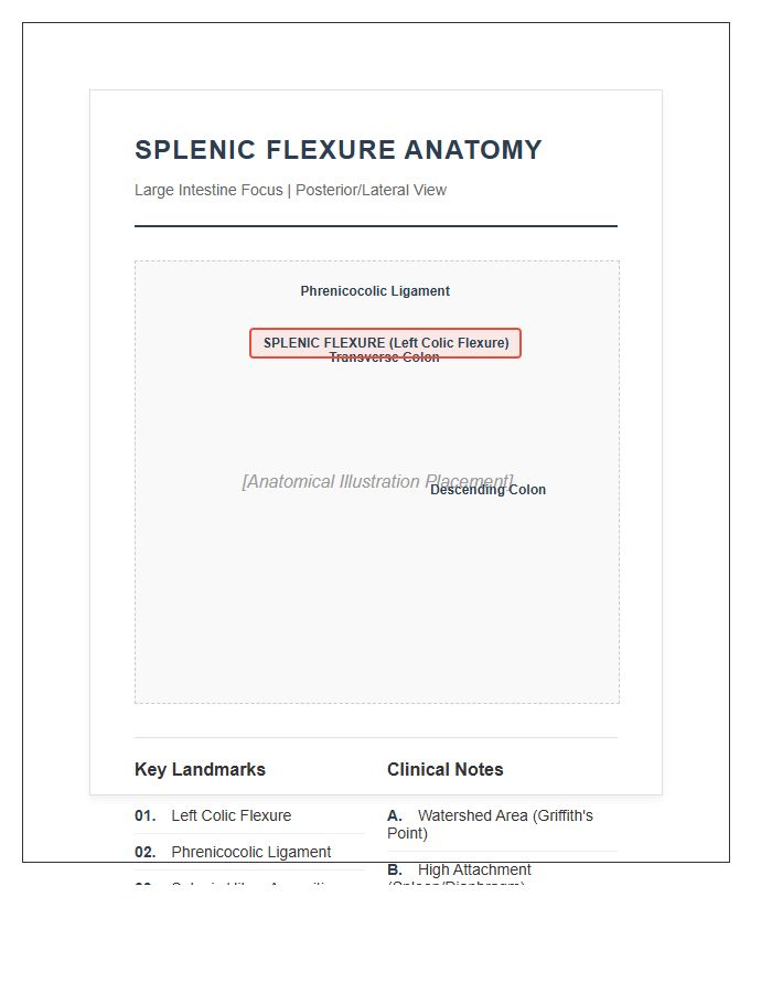
A Splenic Flexure Large Intestine Anatomy Printable Chart is an essential tool for visualizing the sharp bend where the transverse and descending colon meet. It simplifies complex GI anatomy, making it perfect for patient education or exam prep. By clearly mapping this high-pressure zone, these charts help solve common diagnostic confusion and provide a reliable reference for understanding potential bowel obstructions or localized digestive issues quickly.
Large Intestine Muscularis Externa Anatomy Printable Chart
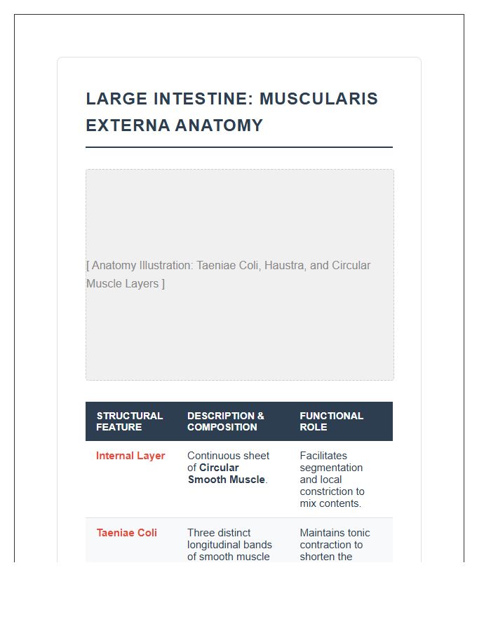
Understanding the Large Intestine Muscularis Externa is essential for mastering gastrointestinal motility. Our printable chart clarifies the unique arrangement of the inner circular layer and the outer longitudinal teniae coli. This visual tool solves the complexity of how muscle contractions move waste efficiently. It is a vital resource for students and professionals needing a quick, reliable reference for digestive anatomy and functional health.
Large Intestine Mesentery Anatomy Printable Chart
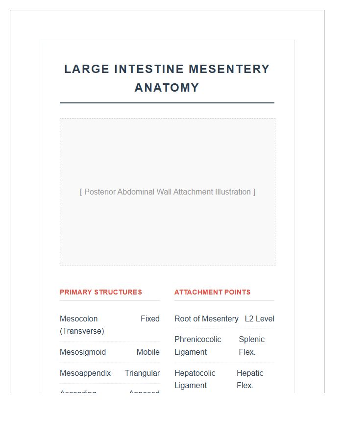
Understanding abdominal health is easier with a Large Intestine Mesentery Anatomy Printable Chart. This visual tool simplifies complex connections, helping you identify structural supports and blood supply routes. It is incredibly useful for students or patients needing to solve anatomical puzzles quickly. Having a high-quality reference ensures you grasp how organs stay anchored, making it an essential resource for clear, effective medical learning.
What are the primary sections of the large intestine included on an anatomy chart?
A detailed large intestine anatomy chart typically illustrates the cecum, the ascending colon, the transverse colon, the descending colon, the sigmoid colon, and the rectum. Each section is distinct in its position and function within the digestive system.
What structural features of the large intestine are visible on a printable diagram?
A high-quality printable chart will show the haustra (small pouches), the taeniae coli (longitudinal muscle bands), and the epiploic appendages (fatty sacs). These unique anatomical markers distinguish the large intestine from the small intestine.
Where is the ileocecal valve located on a large intestine structure map?
The ileocecal valve is located at the junction where the small intestine (ileum) meets the beginning of the large intestine (cecum). On a structural chart, it is found in the lower right quadrant of the abdominal cavity.
What is the function of the hepatic and splenic flexures in intestinal anatomy?
The hepatic flexure is the sharp bend between the ascending and transverse colon on the right side, while the splenic flexure is the turn between the transverse and descending colon on the left. These structures are essential for the directional flow of waste through the abdominal cavity.
How does a printable large intestine chart assist in medical education?
A printable anatomy chart provides a visual reference for identifying the segments, blood supply, and nervous system pathways of the colon. It serves as an essential tool for students and healthcare professionals to study the physiological transition from liquid waste to solid stool.
Note: Sometimes the .PDF file interface can be slightly different from the image preview. Our apology for this inconvenience.

Comments